
Klaus is a 6½-year-old neutered male Wirehaired Pointing Griffon. He is the goofiest dog on earth and is Dr. Green’s personal dog and best buddy. This is a series of dental radiographs taken over a period of 1½ years showing the progression of tooth resorption.
The first sign of tooth resorption was noticed during a routine COHAT (comprehensive oral health assessment and treatment) over 1 ½ years ago.
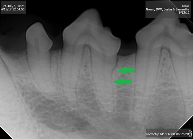
Tooth resorption is a painful and progressive disease with no realistic long-term treatment other than tooth extraction. In early stages with the resorptive lesions well below the gumline, it is not known to be painful and the process can move quickly or very slowly. For several reasons, this tooth was not extracted and we decided to just monitor the tooth annually with dental radiographs under anesthesia.
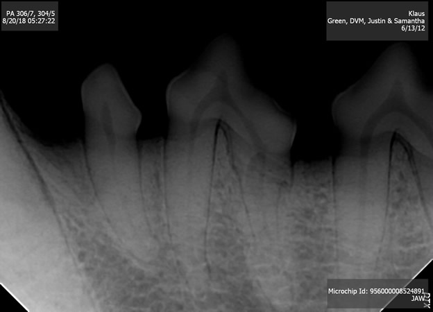
Like all veterinarians working on their own animals, I repeated the radiographs 18 months later rather than 12 months later. The reason for the delay is that all of his teeth looked very clean still and his gums were pink and very healthy. But, Klaus was noticed to consistently avoid chewing on this side of his mouth when he got is daily dental rawhide chew. The following was observed at the follow up COHAT:
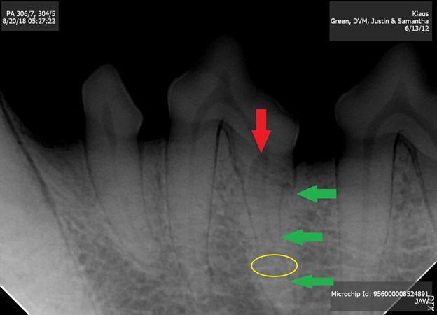
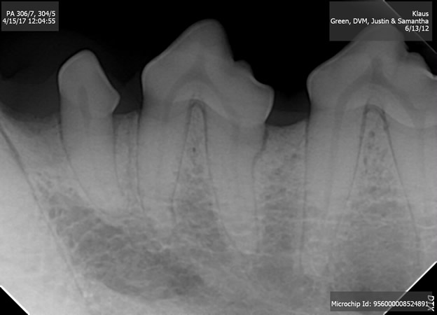
As you can see, the original resorptive area has progressed deeper into the tooth root (green arrows). Additionally, a portion of tooth root near the apex (root tip) has been almost completely resorbed and is now a substance similar to bone (yellow circle). Most importantly, resorption is approaching the crown (red arrow) and this is when the tooth will become painful. The decision was made to extract the tooth.
2 months later when Klaus was brought back for the extraction, the following radiograph was taken immediately before extraction showing even more progression.
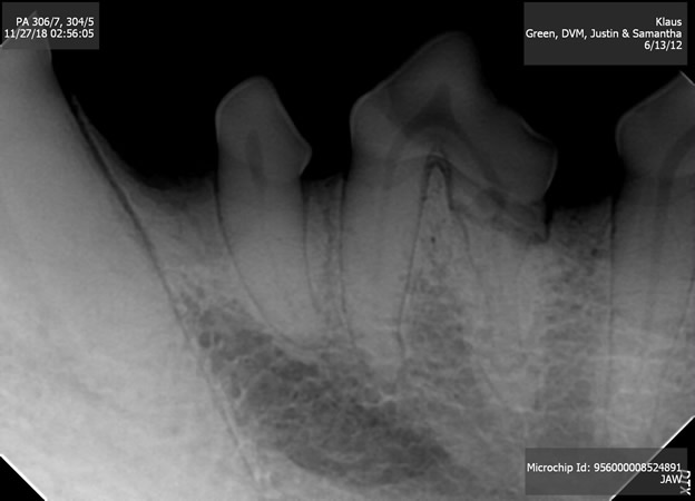
Some of the changes may be caused by a slightly different angle of projection when the radiograph was taken, but these changes are clearly happening quite rapidly.
To extract the tooth, Klaus was placed under general anesthesia with IV fluid administration and full anesthetic monitoring including ECG, blood pressure, pulse-ox and temperature. A breathing tube was placed and his throat was packed with soft gauze to help prevent fluids and debris from irritating his airway.
A regional nerve block was performed with traditional numbing agents (bupivacaine and lidocaine) as well as a narcotic agent called buprenorphine. Adding this micro dose of narcotic to the nerve block may in some cases provide pain control for up to 4 days. This tooth was extracted surgically and very delicately as well as taking post-extraction radiographs to ensure all remnants were removed. Very small absorbable sutures were then placed to close the extraction site and ensure a healthy collar of gingiva remained around the surrounding teeth. He was prescribed anti-inflammatory medication and placed on a soft diet for 14 days and the area healed very nicely.
He already seems to be happier and is chewing on this side of his mouth again. This case is a great example of 3 things:
- A significant, painful dental disease can be occurring below the gum line even with teeth that appear to be very healthy.
- All teeth should be radiographed every time an animal has a dental hygiene procedure. There is no such thing as performing dental x-rays “as needed”.
- The potential dental pain an animal may be experiencing in silence cannot be overestimated. Dogs almost never accurately show their pain and discomfort with dental pain or even most other types of pain. This is why we generally operate on the assumption that if it can cause pain, it is causing pain.
Happy trails!